Most ultrasounds take 15–45 minutes. The time depends on the body area and how many images are needed. You can return to normal activities right after.
Home – Ultrasounds
Professional Radiology offers a full suite of ultrasound imaging services in El Paso. Founded by a world-renowned expert in the field, our clinic is the best in the Southwest for ultrasound scanning services.
Ultrasound scans use high-frequency sound waves to take images, similar to sonar or radar technology. Since ultrasounds use sound waves for imaging, there is no radiation. This is especially ideal when scanning for pregnancy or when diagnosing sensitive conditions. Professional Radiology utilizes the latest in ultrasound technology to support our patients and our city.
Ultrasounds typically don’t require extensive preparation. The only advanced preparations we usually require are a fasting period of 8-12 hours prior to the exam and comfortable clothing. However, some exams, like food or medication restrictions, may require more extensive preparation. Before your exam, our staff will give you a list of pre-exam instructions to follow.
After most ultrasound appointments, you are free to resume normal activities immediately. Ultrasounds are a convenient, safe, and valuable tool for our clinic, our patients, and the doctors we work with.
Professional Radiology offers a variety of ultrasound imaging services to best care for our El Paso patients. Ultrasound technology is probably most associated with pregnancy, but this tech is also used to diagnose issues relating to muscles, tendons, organs, and joints as well.
The following are the services that we offer our patients:
Professional Radiology offers 3D/4D imaging for pregnancy. Through ultrasound technology, we can learn about a baby’s development during pregnancy and help find a due date or detect defects. With the latest in imaging technology, we can provide extremely detailed images of the womb and your child.
Ultrasounds are also excellent for diagnosing vascular issues. Detailed vascular imaging can illuminate potential issues and guide later treatment and/or surgery. Ultrasounds can be useful when looking for diseases like atherosclerosis, peripheral arterial disease (PAD), and more.
Ultrasound technology can be used to examine the liver. In fact, we were the first diagnostic imaging center to offer liver elastography in El Paso. This process detects the stiffness of liver tissue, which can be a sign of conditions like fibrosis and fatty liver disease. Liver issues are on the rise in America, so liver elastography can be key to finding issues early.
Shear wave ultrasounds are the most common type of ultrasound and are available at Professional Radiology. Shear wave elastography uses acoustic radiation (high-frequency sound) to propagate shear waves in the tissue, which can be used to locate stiff, damaged tissue.
Strain elastography is an ultrasound that looks for signs of strain in the tissue. Strain elastography uses compression by the transducer to detect abnormalities. The procedure can be used for a wide variety of organs/tissue types, as well as echocardiography. Strain elastography is a promising recent addition to ultrasound technology and we’re proud to offer it at Professional Radiology.
(915) 225-2480
Ultrasound technology is probably most associated with pregnancy, but this technology is also used to diagnose issues relating to muscles, tendons, organs, and joints as well. The ultrasound procedure is quick and painless. A gel is applied to the area being scanned to help the ultrasound imaging. Once the area(s) has been properly scanned, the gel is cleaned off, and you are ready to resume normal activities. On average, ultrasounds take less than 30 minutes to complete. This simple procedure can provide real-time capture of soft tissue and organ function to guide diagnosis or treatment.
Certain procedures also use ultrasound technology. The two types we perform are Ultrasound-Guided Fine Needle Aspiration and Ultrasound-Guided Biopsy. Both techniques use ultrasound guidance to target the area of interest and minimize risk precisely, but a biopsy generally provides more extensive diagnostic information than a fine needle aspiration.
This is a minimally invasive procedure that uses ultrasound imaging to guide the collection of tissue samples. The procedure uses a very thin, hollow needle to extract a small sample of cells or fluid from a targeted area. It is commonly used to investigate abnormalities or growths detected during an ultrasound exam, such as nodules in the thyroid, breast, or other organs. The small needle size means there is minimal discomfort and risk of bleeding or infection.
The local anesthesia used for the biopsy will usually last for 1-2 hours after the procedure. When the effect wears off, you may have some mild, localized soreness and tenderness at the biopsy sites over the next day or two. You may find regular Tylenol or Ibuprofen is helpful for discomfort. DO NOT TAKE ASPIRIN or MOTRIN twenty-four to forty-eight hours after the biopsy.
If you do experience significant bleeding, redness, infection, or other problems, call your doctor’s office or go to the nearest Urgent Care.
An ultrasound-guided biopsy uses a larger needle to extract a small core tissue sample from a targeted area. It is often used to investigate suspected cancers or other serious conditions in organs like the liver, kidney, prostate, or breast. The larger needle size allows for the collection of a more substantial tissue sample compared to a fine needle aspiration. This tissue sample can provide more comprehensive information for diagnosis and treatment planning.
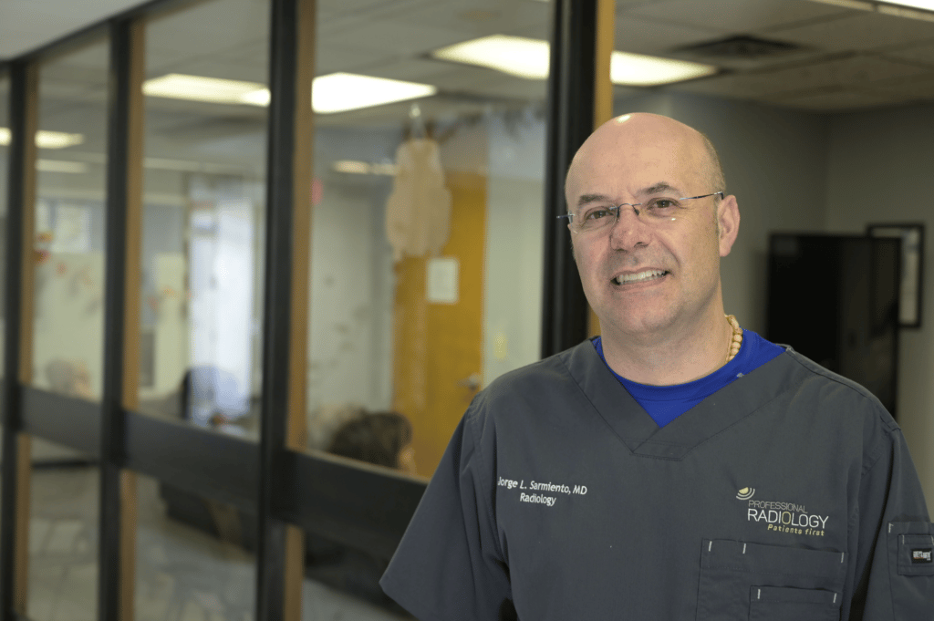
The ultrasound services we offer at Professional Radiology rest on the knowledge and expertise of Dr. Jorge L. Sarmiento, the clinic’s founder and owner. Dr. Sarmiento is a world-renowned radiologist and imaging specialist, with laurels both inside and outside of the clinic.
Dr. Sarmiento has decades of experience in the world of ultrasound. Not only has Dr. Sarmiento been a practicing radiologist for many years, but he has also helped develop 3D ultrasound technology for General Electric. Dr. Sarmiento has also worked as a radiology and ultrasound professor for the UNAM (Universidad Nacional Autonoma de Mexico) and Texas Tech University, respectively. Today, he uses his experience and knowledge to support his patients and staff at Professional Radiology.
A 4D ultrasound shows moving, real-time images of your baby in the womb, creating a video-like picture instead of still images. Many parents enjoy it for bonding and a clearer view.
A KUB (Kidney, Ureter, and Bladder) ultrasound looks at the urinary system. It helps detect kidney stones, blockages, or bladder problems. Doctors use it if a patient is experiencing urinary pain, infections, or blood in the urine.
A TVS (transvaginal scan) is an ultrasound performed with a small probe inserted into the vagina. It provides detailed images of the uterus and ovaries. It’s often used for fertility issues or unexplained pelvic pain.
A Doppler ultrasound measures blood flow through arteries and veins. It helps detect blockages, clots, or circulation problems. Doctors may use it for leg pain, varicose veins, or heart conditions.
The first pregnancy ultrasound usually happens around 6–8 weeks. It confirms the heartbeat and due date. Later ultrasounds monitor growth and development.
The gel helps sound waves travel smoothly from the probe into the body. Without gel, the images would be blurry. It wipes off easily after the scan.
Most ultrasounds take 15–45 minutes. The time depends on the body area and how many images are needed. You can return to normal activities right after.
Easy and convenient scheduling options for our patients.