X-rays and MRI scans are two of the most common diagnostic imaging tests used to evaluate pain, injuries, and other medical conditions. Although both can help doctors see inside the body, they work very differently and are used for different types of problems.
An X-ray is typically faster and especially useful for evaluating bones, fractures, and certain chest conditions. An MRI provides much more detail of soft tissues, including muscles, ligaments, tendons, joints, nerves, the brain, and the spine.
If your doctor is deciding between an X-ray vs. MRI, the right test depends on your symptoms, the area being examined, and the type of injury or condition being considered.
What Is an X-Ray?
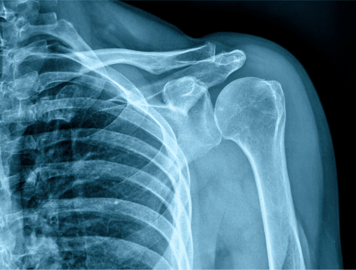
An X-ray is a diagnostic imaging test that uses a small amount of ionizing radiation to create images of structures inside the body.
X-rays are particularly effective for viewing dense structures such as bones.
Doctors commonly order X-rays to evaluate:
- Broken bones
- Joint injuries
- Arthritis
- Bone alignment
- Chest conditions
- Certain infections
- Dental problems
- Some causes of pain
X-rays are widely available, quick to perform, and often used as a first imaging test when a bone injury is suspected.
What Is an MRI?
MRI stands for magnetic resonance imaging.
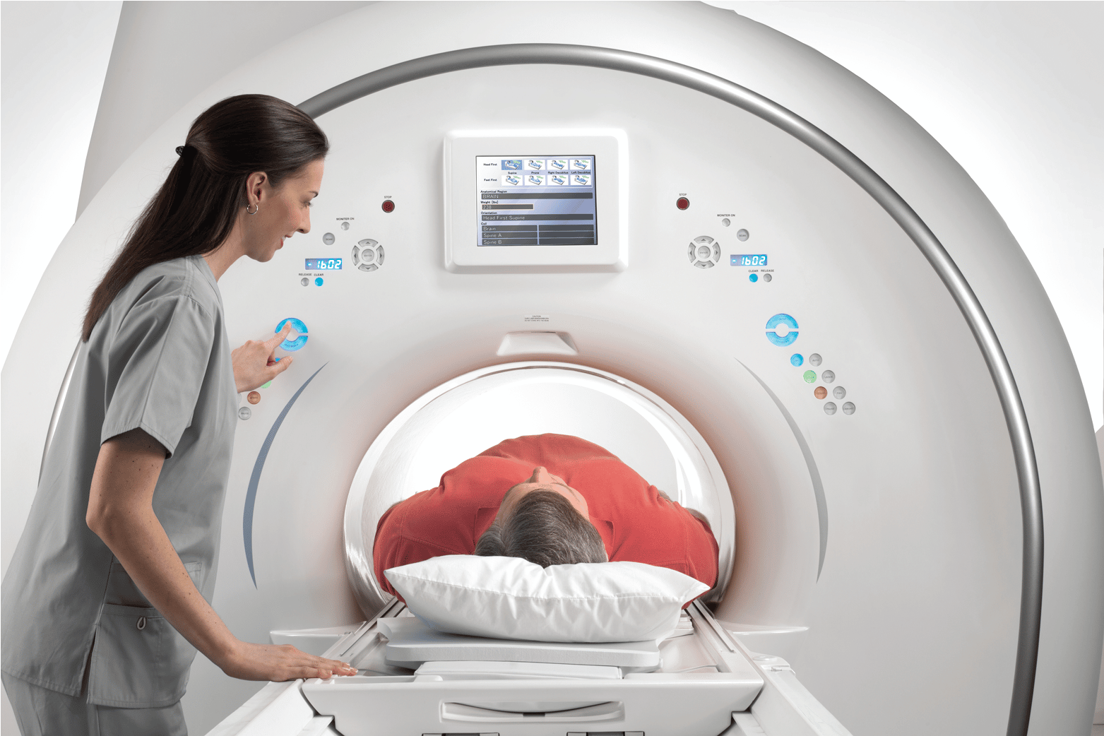
An MRI uses a strong magnetic field and radio waves to create detailed images of organs, tissues, joints, and other structures inside the body.
Unlike an X-ray, MRI does not use ionizing radiation.
MRI scans are especially useful for examining:
- Muscles
- Ligaments
- Tendons
- Cartilage
- The brain
- The spinal cord
- Nerves
- Joints
- Internal organs
- Soft tissue injuries
Because MRI produces detailed images of soft tissue, it may be ordered when an X-ray does not provide enough information.
X-Ray vs. MRI: What Is the Main Difference?
The main difference between an X-ray and MRI is the type of structures each test shows best.
X-rays are highly effective for examining bones and certain dense structures.
MRI scans provide much greater detail of soft tissues.
Another major difference is how the images are created.
X-rays use ionizing radiation, while MRI uses magnetic fields and radio waves.
Which Is Better: X-Ray or MRI?
Neither test is better in every situation.
An X-ray may be the best choice when the concern involves:
- A suspected fracture
- Bone alignment
- Certain joint changes
- Chest symptoms
- Some forms of arthritis
An MRI may be better when the doctor needs to examine:
- Ligaments
- Tendons
- Muscles
- Cartilage
- Nerves
- The spinal cord
- Brain tissue
- Complex joint injuries
Doctors choose imaging tests based on the medical question they are trying to answer.
X-Ray vs. MRI for Broken Bones
X-rays are usually one of the first imaging tests ordered when a broken bone is suspected.
They can help identify:
- Fractures
- Bone displacement
- Joint dislocations
- Changes in bone alignment
However, some small or complex fractures may not always be immediately visible on a standard X-ray.
In certain situations, a doctor may recommend an MRI or another imaging test if symptoms continue despite normal X-ray results.
Can an MRI Detect a Fracture?
Yes. MRI can detect certain fractures and bone injuries.
MRI may be particularly useful for identifying stress fractures, bone marrow changes, or injuries that are difficult to see on a standard X-ray.
However, MRI is usually not the first test ordered for a straightforward suspected fracture.
X-Ray vs. MRI for Knee Pain
Both X-rays and MRI scans may be used to evaluate knee pain, but they provide different information.
A knee X-ray can help evaluate:
- Bone injuries
- Arthritis
- Joint spacing
- Bone alignment
A knee MRI provides detailed images of:
- Meniscus
- ACL and other ligaments
- Cartilage
- Tendons
- Muscles
- Other soft tissues
If a doctor suspects a ligament tear or meniscus injury, an MRI may be more useful than an X-ray.
X-Ray vs. MRI for Back Pain
Imaging is not always necessary for back pain, but when diagnostic imaging is appropriate, the test depends on the suspected cause.
An X-ray may help evaluate:
- Bone alignment
- Certain fractures
- Degenerative changes
- Some structural abnormalities
An MRI can provide detailed images of:
- Spinal discs
- Nerves
- Spinal cord
- Ligaments
- Soft tissues
MRI may be recommended when symptoms suggest a herniated disc, nerve compression, spinal cord problem, or another condition involving soft tissue.
X-Ray vs. MRI for Shoulder Pain
An X-ray may be used as an initial imaging test for shoulder pain because it can show fractures, arthritis, and certain bone abnormalities.
An MRI may be recommended when doctors suspect:
- Rotator cuff tears
- Tendon injuries
- Labral tears
- Ligament injuries
- Cartilage damage
The test your doctor orders depends on your symptoms and physical examination.
X-Ray vs. MRI for Neck Pain
For some patients with neck pain, an X-ray may provide enough information to evaluate bone alignment or degenerative changes.
An MRI may be recommended when the concern involves:
- Nerve compression
- Herniated discs
- Spinal cord problems
- Persistent neurological symptoms
- Soft tissue injuries
Symptoms such as weakness, numbness, or tingling may sometimes lead a healthcare provider to consider more detailed imaging.
X-Ray vs. MRI for Ankle and Foot Injuries
X-rays are commonly used after ankle and foot injuries when a fracture is suspected.
If the X-ray is normal but pain continues, an MRI may be useful for evaluating:
- Tendon injuries
- Ligament tears
- Stress fractures
- Cartilage damage
- Soft tissue injuries
The appropriate imaging test depends on the type and severity of the injury.
X-Ray vs. MRI for Hip Pain
Hip X-rays can help identify conditions involving bones and joints, including certain fractures and arthritis.
MRI may provide additional information about:
- Muscles
- Tendons
- Cartilage
- Bone marrow
- Labrum
- Stress injuries
Your doctor may start with an X-ray and recommend an MRI if more detail is needed.
Can an X-Ray Show Ligament Damage?
Standard X-rays do not directly show ligaments in the same detail as MRI.
An X-ray may show changes in bone alignment or other findings that suggest an injury, but MRI is generally much better for evaluating ligament tears.
This is one reason MRI is commonly used for sports injuries involving the knee, shoulder, ankle, or other joints.
Can an X-Ray Show Muscle Injuries?
X-rays are not designed to show muscles in detail.
If a doctor suspects a significant muscle, tendon, or other soft tissue injury, MRI or another imaging test may be more appropriate.
Can an MRI Show Bones?
Yes. MRI can show bones, but it visualizes them differently than an X-ray.
MRI is especially useful for looking at:
- Bone marrow
- Stress injuries
- Certain fractures
- Tumors
- Infections
- Surrounding soft tissues
For straightforward bone fractures, X-rays are often faster and more practical.
Which Imaging Test Is Better for Arthritis?
X-rays are commonly used to evaluate arthritis because they can show changes in joint spacing, bone structure, and certain degenerative changes.
MRI can provide more detailed images of cartilage, ligaments, bone marrow, and other joint structures.
A doctor may order an MRI if symptoms cannot be fully explained by X-ray findings or if more detailed information is needed.
X-Ray vs. MRI for Sports Injuries
The right imaging test for a sports injury depends on what may be damaged.
An X-ray is often used first if a broken bone is suspected.
MRI is commonly used for injuries involving:
- ACL tears
- Meniscus tears
- Rotator cuff injuries
- Tendon tears
- Ligament sprains
- Cartilage damage
- Muscle injuries
- Stress fractures
MRI can provide detailed images of structures that are difficult to evaluate with X-rays.
Does an X-Ray Use Radiation?
Yes.
X-rays use ionizing radiation to create images.
The amount of radiation depends on the type and number of X-rays performed.
Healthcare providers consider the medical benefit of imaging when deciding whether an X-ray is appropriate.
Does an MRI Use Radiation?
No.
MRI does not use ionizing radiation.
Instead, it uses a strong magnetic field and radiofrequency energy to produce detailed images.
This makes MRI different from X-ray and CT imaging.
Which Test Is Faster: X-Ray or MRI?
An X-ray is generally much faster than an MRI.
The imaging portion of an X-ray may take only a few minutes.
MRI scans often take approximately 20 to 60 minutes, depending on the body part being examined and the complexity of the test.
Which Test Is More Comfortable?
X-rays are usually quick and require minimal time in a fixed position.
MRI exams require patients to lie still for longer periods.
Some people may also feel uncomfortable in an MRI scanner because of:
- Limited space
- Loud noises
- Claustrophobia
- The need to remain still
If you are concerned about claustrophobia, tell the imaging center before your MRI appointment.
Does MRI Require Contrast?
Not every MRI requires contrast.
Some MRI scans use contrast material to make certain tissues, blood vessels, or abnormalities easier to see.
Whether contrast is needed depends on the area being examined and the medical question.
Standard X-rays generally do not require contrast, although some specialized X-ray procedures may use contrast agents.
Is an MRI More Accurate Than an X-Ray?
MRI is not automatically more accurate than an X-ray.
The better test depends on what needs to be evaluated.
For example, an X-ray may provide exactly the information needed to diagnose a straightforward fracture.
MRI may be much more informative when doctors need to evaluate ligaments, nerves, cartilage, or other soft tissues.
Using the correct imaging test is more important than choosing the test that produces the most detailed image.
Why Would a Doctor Order an X-Ray Before an MRI?
Doctors often begin with an X-ray because it is:
- Fast
- Widely available
- Effective for evaluating bones
- Useful for identifying common causes of pain
- Often enough to answer the initial medical question
If the X-ray does not explain the patient’s symptoms, an MRI may be ordered for a more detailed evaluation.
Why Would You Need an MRI After a Normal X-Ray?
A normal X-ray does not rule out every type of injury.
An MRI may be recommended after a normal X-ray if your doctor suspects:
- Ligament damage
- Tendon injury
- Cartilage damage
- Herniated discs
- Nerve compression
- Stress fractures
- Other soft tissue abnormalities
MRI can detect problems that may not be visible on standard X-rays.
What Symptoms May Lead to an X-Ray?
A healthcare provider may consider an X-ray if you have symptoms or injuries involving:
- Sudden pain after an accident
- Swelling
- Difficulty moving a joint
- Suspected broken bones
- Chest symptoms
- Joint pain associated with arthritis
- Bone deformity
Your doctor will determine whether imaging is necessary based on your symptoms and physical examination.
What Symptoms May Lead to an MRI?
MRI may be considered when symptoms suggest problems involving nerves or soft tissues.
Examples may include:
- Persistent joint pain
- Muscle weakness
- Numbness
- Tingling
- Suspected ligament tears
- Suspected tendon injuries
- Neurological symptoms
- Persistent back or neck pain with certain warning signs
The decision to order an MRI depends on the overall clinical situation.
How Do You Prepare for an X-Ray?
Most standard X-rays require very little preparation.
You may be asked to:
- Remove jewelry
- Remove metal objects
- Change into a medical gown
- Tell the technologist if you may be pregnant
Depending on the area being examined, additional instructions may be provided.
How Do You Prepare for an MRI?
MRI preparation requires additional safety screening because of the strong magnetic field.
Before your exam, tell the imaging center about:
- Pacemakers
- Metal implants
- Cochlear implants
- Joint replacements
- Previous surgeries
- Metal fragments
- Implanted medical devices
- Pregnancy
- Claustrophobia
You will also need to remove metal objects before entering the MRI room.
Frequently Asked Questions About X-Ray vs. MRI
Is an MRI better than an X-ray for joint pain?
It depends on the suspected cause. X-rays are useful for bone and joint changes, while MRI provides more detailed images of cartilage, ligaments, tendons, and other soft tissues.
Should you get an X-ray or MRI for a knee injury?
An X-ray may be used first if a fracture is suspected. MRI may be recommended for suspected ligament, meniscus, tendon, or cartilage injuries.
Can an MRI see things an X-ray cannot?
Yes. MRI can provide detailed images of nerves, muscles, ligaments, tendons, cartilage, and other soft tissues that are not clearly visible on standard X-rays.
Is an MRI safer than an X-ray?
MRI does not use ionizing radiation. X-rays do use radiation, although they are commonly performed when medically appropriate. MRI also has its own safety considerations because of its strong magnetic field.
Why not get an MRI instead of an X-ray every time?
MRI takes longer, is more complex, and may provide information that is unnecessary for certain conditions. A standard X-ray may be the most appropriate and efficient test for many bone-related problems.
X-Ray and MRI Services in El Paso, TX
Choosing between an X-ray and MRI depends on the symptoms you are experiencing and the structures your healthcare provider needs to evaluate.
X-rays are often an excellent first choice for fractures and bone-related conditions, while MRI provides detailed imaging of soft tissues, joints, nerves, the brain, and the spine.
Professional Radiology provides digital X-ray and MRI services in El Paso, TX, along with additional diagnostic imaging services for patients and referring healthcare providers.
If your doctor has ordered an X-ray or MRI in El Paso, contact Professional Radiology to learn more about scheduling your diagnostic imaging exam.