Have you ever wondered, “How did they ever think of that?” The truth is, most scientific breakthroughs are made only after extensive experimentation with gradual gains, with each researcher building on the work of those who came before.
But many of the world’s greatest inventions were based on discoveries made by accident. Penicillin, microwaves, Coca-Cola, and Super Glue are just a few examples. But few accidental discoveries (with the possible exception of penicillin) have had the impact on humanity that the X-ray has. And it has a fascinating history.
The Discovery of the X-Ray
In 1895, Wilhelm Roentgen, a professor of physics in Bavaria, was working on an experiment with cathode ray tubes to learn if cathode rays could travel through a vacuum tube. He applied a high voltage to the tube and noticed that the positive and negative electrodes within the tube caused it to emit light.
He then covered the tube with black paper to see if the light would shine through, and a nearby screen treated with a chemical called barium platinocyanide began to glow.
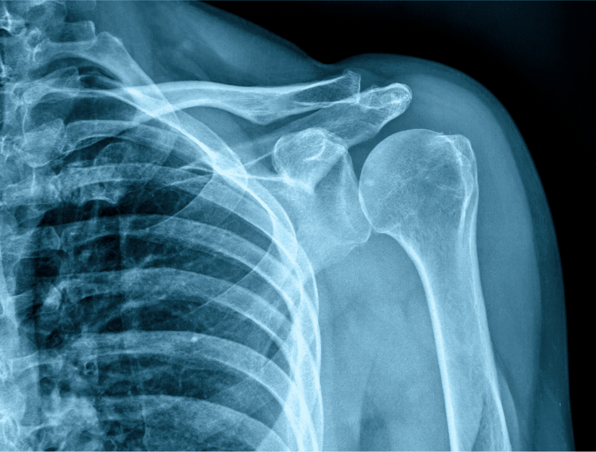
He concluded that a type of radiation must be at work from inside the tube. He soon learned that the rays had very short wavelengths that enabled them to pass through human flesh and leave a shadow of the underlying bones on the screen.
He named this radiation X-ray, with X standing for unknown. He created the very first X-ray by capturing the image of the bones of his wife’s hand. When she looked at the image, his wife is said to have cried, “I have seen my death!”
For his discovery of radiation, Roentgen won the very first Nobel Prize in physics in 1901. But he couldn’t have known that his discovery would become one of the cornerstones of modern medicine.
X-Ray’s Early Contribution to Medicine
A year after Roentgen’s discovery, in 1896, Dr. Edwin Frost and his brother, Dr. Gilman Frost, were the first to take a diagnostic X-ray. They X-rayed a boy named Eddie McCarthy to diagnose a broken wrist.
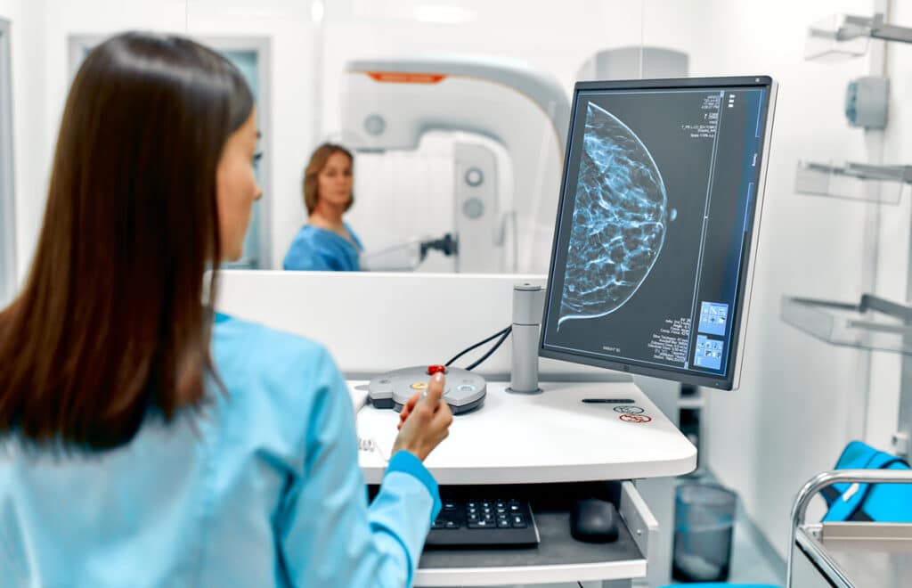
Also in 1896, Emil Grubbe of Chicago is thought to be the first to use radiation to treat cancer; he successfully treated a woman with breast cancer. (Grubbe would go on to die in 1960 from cancer due to radiation exposure.)
Around this same time, Thomas Edison started his own X-ray research. He experimented with various materials to learn which was the most effective to use for the screen. He settled on calcium tungstate, and with it, he developed the first mass-produced imaging device, the “Vitascope.” Today we call his invention the fluoroscope.
In 1913, William Coolidge invented the Coolidge vacuum tube. These tubes create continuous X-ray emissions, are more stable than the cathode ray tubes, and allow the intensity and energy of the rays to be controlled separately. The Coolidge gradually replaced the cold cathode-ray tubes, and this design is still in use today.
Discovering the Dangers of X-Ray
It was during Edison’s research that the dangers of X-rays were discovered. Clarence Dally, a glassblower who worked with Edison, would X-ray his own hands to test X-ray tubes. Eventually, he had both of his arms amputated due to cancer, and he died of X-ray exposure in 1904. From then on, Edison wasn’t fond of the technology and stopped his research. “Don’t talk to me about X-rays,” he famously said. “I am afraid of them.”
Dr. D.W. Gage of Nebraska was also discovering the dangers of radiation exposure at that time, including reddened skin, skin lesions, skin sloughing off, and hair loss.
X-Ray Advancements
Refinements and advances in equipment design since 1920 have made X-rays much safer for patients and technicians.
Today’s films use chemicals that make them more sensitive to X-rays, so they require less time and less radiation to create an image. The most sensitive of these screens use rare-earth metals.
All radiology technicians now wear lead aprons when taking X-rays, which the rays can’t penetrate.
Medical providers are more careful now to weigh the benefit versus risk before ordering X-rays to limit the long-term cumulative effect of radiation on patients.
Beyond the X-Ray
Interestingly, when word traveled about the x-ray back in 1895, it created such a sensation that people were using it more for photographic than medical purposes. Anyone could create a cathode ray tube and take photos with it. And many people did.
Photographers set up on the street to take X-rays of passersby. Shoe merchants even used an X-ray machine to aid in the fitting of shoes.
But once people realized the medical applications of the X-ray, its use spread quickly. With the Coolidge tube, X-ray became the gold standard for diagnosing fractures, foreign bodies, pneumonia, and many other ailments.
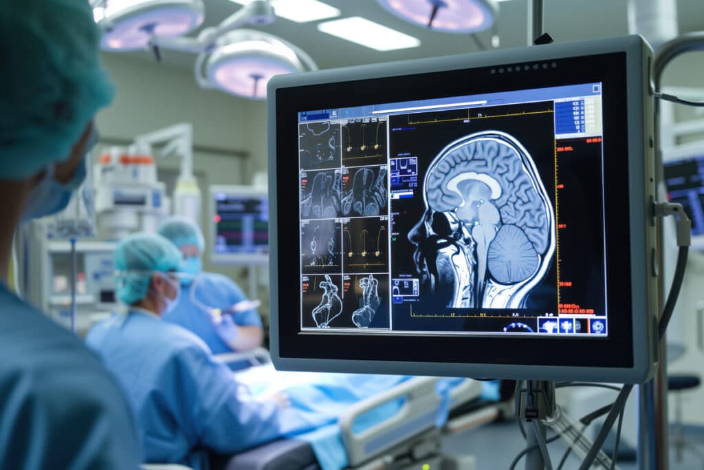
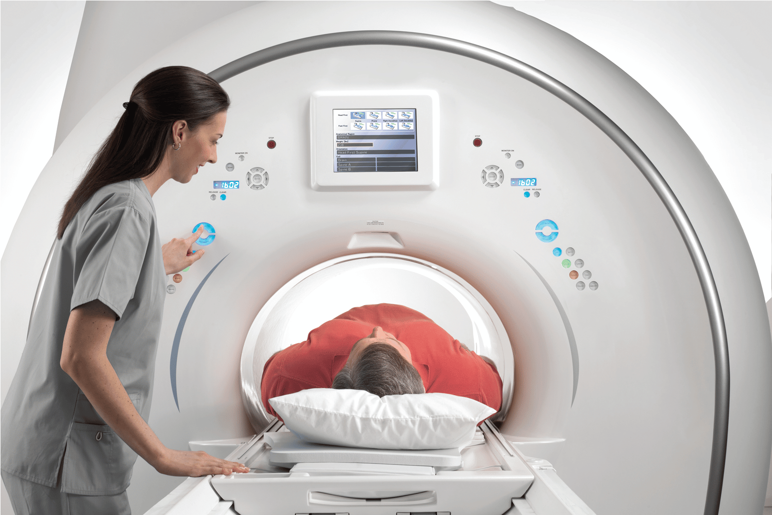
As the field of radiology has grown, X-rays and fluoroscopy led the way to more sophisticated and safer imaging techniques. These include CT (or CAT) scan, MRI, electrocardiography, ultrasound, and nuclear imaging (such as the PET scan), some of which don’t use radiation at all. Not bad for a short wave of light discovered by accident!
If you’d like to experience the groundbreaking benefits of x-rays, then contact Professional Radiology!