The technology behind elastography has incredible applications for disease diagnosis in the body. If your doctor suspects you may have serious issues with your liver, kidney, prostate, or other gland or organ, then it’s likely for them to order an elastography test.
Unfortunately, many people simply aren’t aware of what elastography is, and just how crucial it can be in getting accurate care. For healthcare professionals, this test has been a life-saving tool for care since the invention of technologies such as ultrasound and MRI. We’ll briefly take you through the history, uses, and breakthroughs of the practice.
What Exactly Is Elastography?
Elastography, in simple terms, is a medical imaging technique to map how stiff or elastic soft tissue is. In most cases, your soft tissue will be soft and elastic. Diseased or cancerous tissue, on the other hand, will be much harder and/or stiffer. Think about a freshly chewed piece of gum versus an old, dried out piece. Using this example, elastography will map out the ratio of fresh gum and dry gum in certain parts of your body.
More specifically, however, elastography works by sending low-frequency vibrations through an area and observing how the tissue reacts. The vibration is different depending on the type of elastography, but the basic idea is to use distortions to map the area. The two most used methods of distortion are strain and shear wave. Strain elastography uses pressure to map tissue displacement (how much it moves) while the shear wave method sends waves through the tissue and measures the speed of the waves.
But how do vibrations lead to these measurements? We’re glad you asked! Stiff/hard tissue responds differently to the force. The pressure or waves that are produced by the distortion actually move faster through the stiff tissue and deforms less.
Elastography is split into different types based on the tech used. Ultrasound and magnetic resonance imaging are some of the most common techniques, but there are several other methods that can be used. The technology is also categorized by the dimensions in which the data is shown (1D, 2D, 3D). Some technologies will be used together to form the most accurate image possible.
Needless to say, it would be a little harder to accurately diagnose and pinpoint diseases without this technology. But doctors still had to do this work before the invention of this awesome technology. Now we’ll take a brief look at the history of the practice.
History of Elastography
For the majority of the 20th century, the idea of elastography using machinery was all but ignored. Palpation, the precursor to elastography, required the use of a doctor’s hands to feel for abnormalities and has a history dating back several millennia. The inherent issue with this method is that it requires a human interpretation of what feels stiff and abnormal, rather than concrete imaging like we have today. In the 2oth century, a few papers here and there discussed vibrations and soft tissue, but the scientific community simply wasn’t all that interested in it.
The first real mention of elastography in the way it’s used today was in the 1960s, but most early research focused on the acoustic assessment of skin, not other soft tissue. It wouldn’t be until the ‘80s and ‘90s that the idea of using waves to investigate other forms of tissue would take shape.
It wouldn’t be until the early 2000s that the first reliable machines would be introduced into the medical world, forever changing the way many diseases were documented and investigated. Now there are several different types of elastography technology that radiologists can choose from to accurately diagnose a patient.
What Elastography Helps Diagnose
As stated before, the first research on the efficacy of elastography focused on the skin, rather than internal tissue. The first use of elastography for internal tissues was in France, where they used the ground-breaking technology on the livers of several patients. To date, the liver is one of the most investigated organs using elastography.
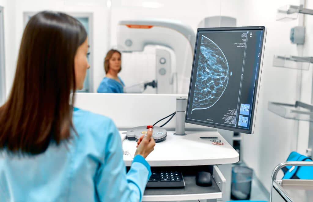
Elastography’s uses are actually quite broad. Skin, internal tissues, and even musculoskeletal imaging are all made possible with this technology. Organs like the thyroid, prostate, and breasts are all commonly investigated using this elastography. In fact, many cancers and diseases have been detected using this new tech. If you have serious health complications, there’s a big chance you’ll be diagnosed using some of this life-saving technology.
Expert Care From Professional Radiology
As much as elastography has completely changed medical care, it still takes a discerning eye and hand to find the right diagnoses. In fact, Professional Radiology is one of the few imaging centers in El Paso with the technology and expertise necessary to produce the best results. The Professional Radiology team represents El Paso’s best radiology experts, providing expert care for every client we work with. Give us a call today to learn more about how we can help!