In the early 1990s, the pink-colored ribbon began its cultural trajectory of representing the struggle, survival, and incredible resilience of women fighting against breast cancer. The color pink was established for the movement and the ribbon was the symbol.
October is a month to celebrate the victories, remember the struggles, and learn about the fight against breast cancer. Tucked away beneath all of the symbolism and public events, is the quiet experience of the mammogram and the radiology technology that makes it possible. Breast cancer has been around for a long time, but the technology and knowledge we currently use to combat it have an incredible history.
Why are mammograms at the center of this public health battle?
Here’s what you need to know.
What is Breast Cancer?
This cancer is characterized by the growth of malignant cells in the epithelial tissue of the breast. As defined by Harvard Health, abnormal growth can occur in various areas of the breast including the ducts that carry milk to the nipple, the milk-producing sacs, and other non-glandular tissue.
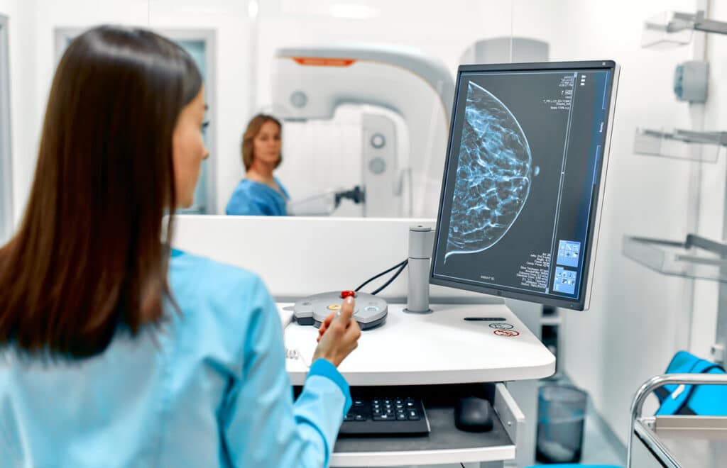
The advent of mammography remains the go-to for breast imaging. This is a subspecialty area of radiology that specializes in the images of breast tissue for the purpose of detecting cancer. The field has—like every other area in medicine—advanced significantly since its humble beginnings. Especially in the area of precise imaging, the work done by breast imaging radiologists is essential for women’s health today.
Mammography screenings became a standard of care after randomized and controlled trials showed how essential early detection is in order to decrease mortality rates from breast cancer.
Why is a Mammogram Important?
The short answer is detection. Early detection can save lives.
The longer answer is that despite a lack of breast cancer in your family, screening is vital as 75% of women diagnosed with breast cancer have no cited family history. Breast cancer often has no symptoms in its early stages. Annual screenings are recommended by the American College of Radiology for women over 40. The American Cancer Society has reported that early-stage breast cancer has a five-year survival rate of 99 percent.
Breast cancer death rates have gone down 40% since 1989. Several factors can be at the center of this including optimal treatment, improved detection, more precise screening tools, and technology, and increased awareness.
What to Expect From Your First Mammogram
The idea of getting the screening can be stressful or a little intimidating. Today’s techniques, however, are quite effective, and relatively painless. The whole process takes about thirty minutes!
Keep in mind:
- Suspicious findings are not always cancers, many women can have cysts, dense tissue, or an unclear image
- Wear a two-piece outfit to make it easier. This way you only have to remove the top garments.
- Each of the breasts will be compressed for about half a minute. The process of compression might be uncomfortable but it is an important aspect of getting the best image.
A Quick Timeline and Trajectory of the Radiology Technology
Advancements in the area of imaging and film contributed to the improvement of mammography techniques. A deep dive into the radiology techniques can get technical pretty quickly, but the place where we are today is a compounded result of many dedicated individuals, physicians, and scientists that fought for better imaging technology through the decades.
- In the 1960s, mammography exams used general-purpose X-ray tubes without compression. The images that resulted had little contrast and some underexposed areas simply appeared white.
- In the 1970s, screen-film mammography was possible with a lower radiation dose and provided greater visibility through the brain tissue.
- In 1976, the American Cancer Society recommends mammography as a detection tool
- As breast cancer was categorized as a public health threat, regulations and consistency in the use of radiology in mammography screenings became a priority
Mammography at the Turn of the 21st Century
Breast imaging continues to become more effective and useful in detecting early breast cancer. As the digital revolution hit every industry imaginable, it also became a major factor in radiology. Digital mammography works similarly to analog mammography. The main difference is that the end result is in digital information rather than X-ray film.
The positive results of digital mammography have improved detection capabilities. This newer technology can:
- Provide more precise results for dense breasts or those of younger patients
- Use a lower radiation dose than older technologies
- Radiologists can focus on abnormalities in women with dense breasts
The findings regarding improved detection rates for women with dense breasts were published in the New England Journal of Medicine, after a large clinical study that compared the use of digital mammography with traditional. The reason for this, as outlined by Harvard Health Journal, is that dense breast tissue has less fat and more glandular and connective tissue.
Nevertheless, traditional mammography proved just as effective as digital mammography in women who did not have dense breasts. The same clinical study showed that digital technology was just as effective as the traditional when it came to non-dense breast tissue.
Embrace Breast Cancer Awareness Month and Find Out if It’s Time to Schedule a Mammogram!
Early detection saves lives. This is one of the mantras of breast cancer awareness advocates because it is statistically true. Mammography is a marvel of imaging and modern technology. And the month of October is Breast Cancer Awareness Month, so why not learn more about your risks and what you can do to stay healthy.
Have questions about the mammogram procedure and what to expect? Contact Pro Radiology to learn more.