Diagnostic imaging tests are tools used by physicians to diagnose a range of medical conditions. Each of these imaging methods uses different technologies to create real-time images and videos of the internal structures of the body. It helps doctors narrow down the possible causes of an illness or pain to help them find out the accurate diagnosis.
There is no need to be anxious as imaging tests are non-invasive and painless. For X-rays, it usually takes less than 10 minutes. An ultrasound can take about 30 minutes to an hour while CT scans can take 10-20 minutes. The magnetic resonance imaging may take longer, depending on the area being scanned. It can take up to 90 minutes, max.
Why Diagnostic Imaging Methods Are Important
Medical imaging is used to diagnose, monitor, and treat medical problems. They are crucial for detecting various diseases such as cancer. With medical imaging, doctors can detect a tumor at an earlier stage, reducing deaths by about 20 percent. In some cases, early detection of other diseases can potentially save a life.
This article will talk about the different diagnostic imaging methods such as X-rays, CT scans, Ultrasound, and MRI.
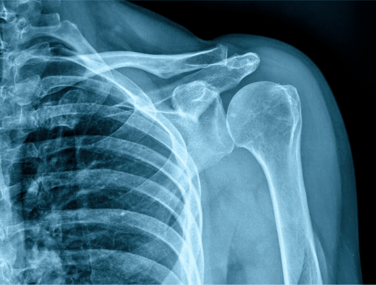
X-ray
Also called a radiograph, an X-ray uses radiation to create images of the body. It’s the fastest and the most common form of diagnostic imaging. X-rays are often used to diagnose problems or complications with the bones. Patients that suffered fractures, misalignments, or dislocations are usually asked to get an X-ray so the medical doctors can give a proper diagnosis. X-rays are also used to diagnose infections, tumors, and bone disease.
During an X-ray exam, the body part that needs to be checked is placed between the X-ray machine and a photographic film. The machine sends electromagnetic waves through the body, reflecting its internal structures on the film. However, when the rays pass through bones and teeth, they will appear white on the photographic film. Frequent exposure to X-rays can be harmful to the patient, but certain precautions are taken, especially when pregnant.
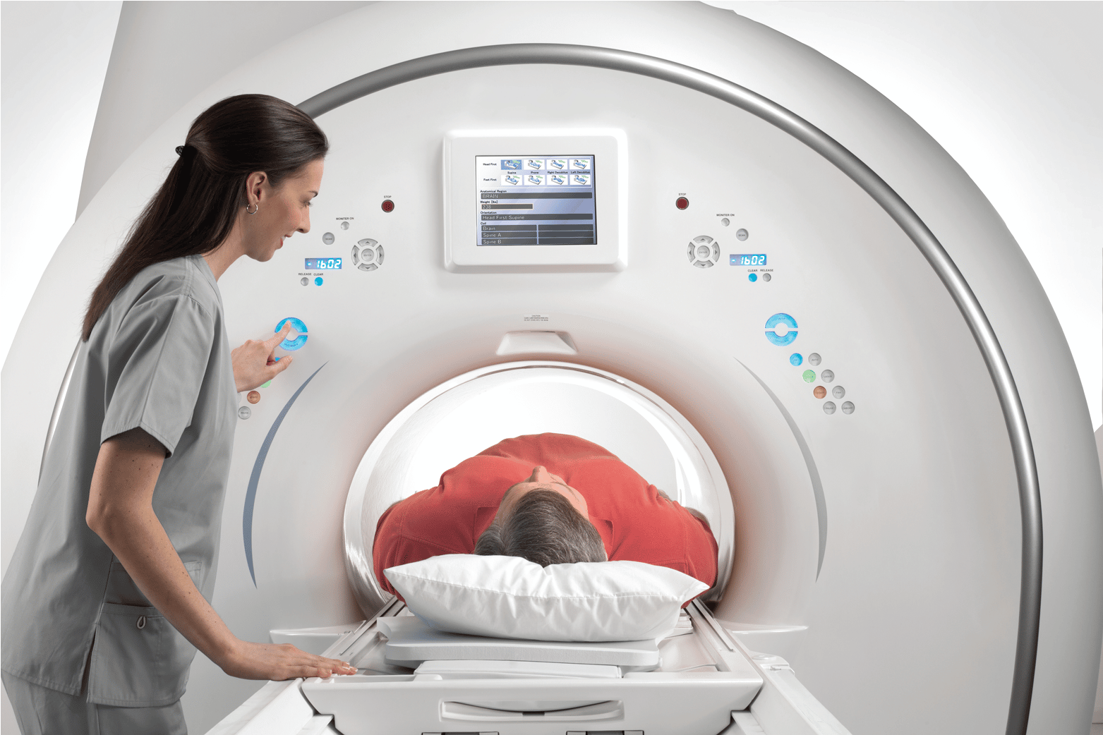
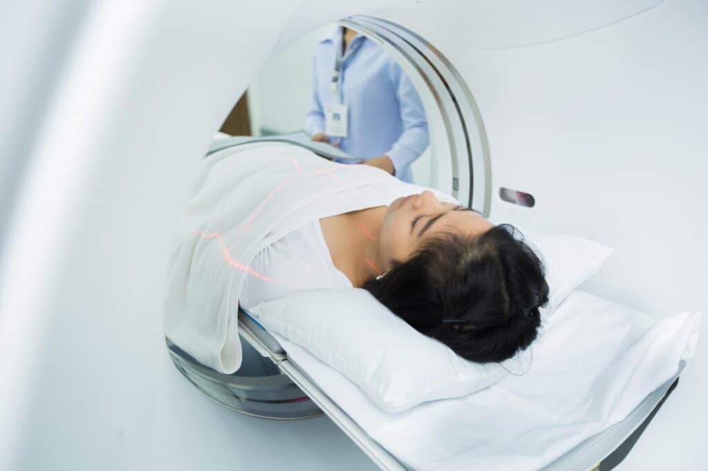
CT Scan
Originally, a CT scan was created to produce detailed pictures of the brain. Today, doctors recommend a CT scan to take photos of the internal organs, blood vessels, soft tissues, and bones. Doctors may order a CT Scan to help them determine the location and exact size of a tumor.
Like the X-ray, a CT scan sends radiation through your body with a much detailed view of its structures. It can produce detailed images of the blood vessels and the body’s different organs and is often used to help diagnose heart disease, cancer, appendicitis, and infectious diseases.
The patient ordered to undergo a CT scan is asked to lie on the CT scanner equipped with a tunnel-like structure in the center. As the scanner rotates, it produces a 360-degree image of the body’s internal organs.
This diagnostic image is more pricey than X-rays or Ultrasounds.
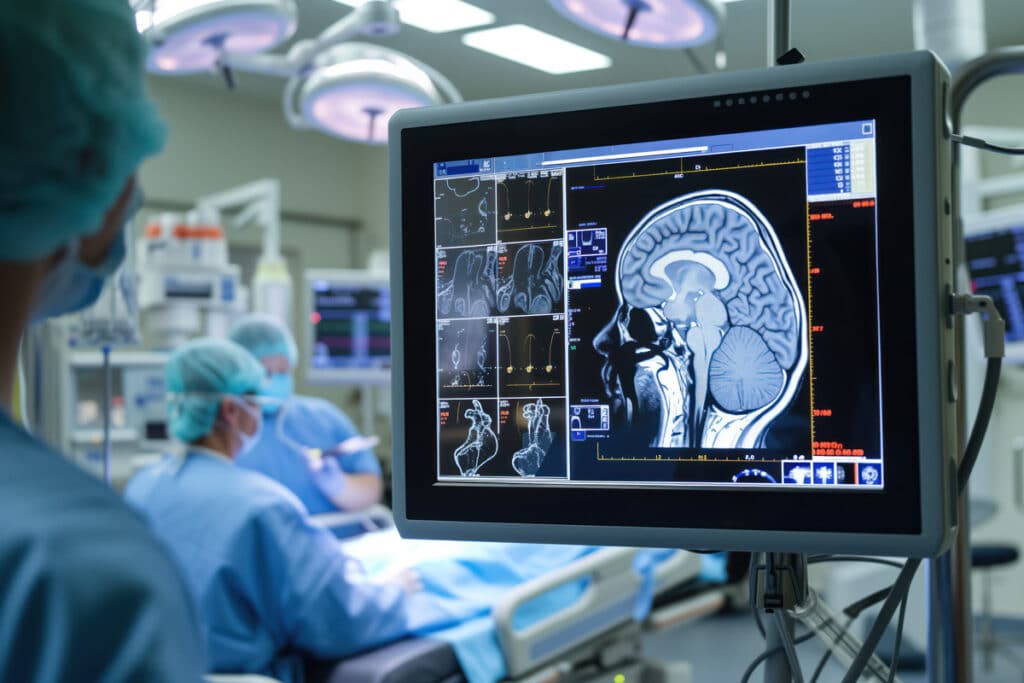
MRI
Magnetic resonance imaging or MRI uses a powerful magnet for passing radio waves through your body to create detailed images of its internal structures. It can detect damaged tissues, abnormalities, and cancerous growths. When getting an MRI, the patient needs to remain very still for the scanner to produce high-quality images. Infants and children may be sedated during the entire procedure.
The doctor often orders an MRI to take detailed images of the body. Compared to a CT scan, an MRI can provide better soft-tissue contrast and differentiate better muscle, water, soft tissue, and fat. Unlike X-rays, MRIs don’t have the harmful effects of radiation. However, it’s equipped with a strong magnetic field that may attract magnetic objects and create loud noises. Patients with implanted devices may be recommended to get a CT scan instead.
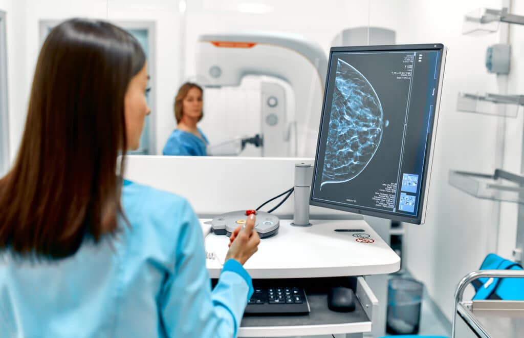
Ultrasound
Also known as sonography, ultrasound technology uses high-frequency sound waves to create a live video feed of the inside of the body. It can show the movement of the internal organs and blood flowing through the blood vessels. Because it doesn’t have any radiation, an ultrasound is the most preferred test for viewing a developing fetus. Surgeons also use ultrasound to aid them during biopsies or other medical procedures. Ultrasound guidance is also used for treating varicose veins.
Your doctor may ask you to get an ultrasound when you’re experiencing swelling, pain, or other symptoms that require an internal view of your organs. If there are abnormalities, the doctor may ask you to undergo further tests such as an MRI or a CT scan.
What to Do When You Have Health Concerns
If you are experiencing any pain or other symptoms, consult with a physician immediately. Diagnostic imaging tests can help diagnose illnesses and other diseases. However, you do need to speak with a doctor first for a medical checkup. During a doctor’s visit, you may be ordered to have a diagnostic exam, and the test depends on your symptoms. Further diagnostic exams may be ordered when the doctor sees certain abnormalities in the results.
Get in Touch With Dr. Jorge L Sarmiento at Professional Radiology
Do you need to undergo CT scans or X-rays? If you need medical diagnostic imaging in El Paso, don’t hesitate to get in touch with us. Dr. Jorge L. Sarmiento has decades of experience and hands-on training in the art of medical imaging. Professional Radiology is a physician-owned clinic committed to providing accurate and high-quality imaging services to residents of El Paso and the surrounding area. Our goal is to offer compassionate care by intently listening to our patients. Our courteous and professional staff aims to provide the best care for anyone needing imaging tests in El Paso. Contact us today for an appointment.